
Cognition Neuroimaging
Cognition Neuroimaging Neuro imaging in many respects revolutionized the study of cognitive neuroscience, the discipline that attempts to determine the neural mechanisms underlying cognitive processes. Early studies of Brain-behavior relationships relied on a precise neurological exam in the basis for hypothesizing the site of brain damage that was responsible for a given behavioral syndrome. The advent of structural brain imaging, first with computerized tomography and later with magnetic resonance imaging, paved the way for more precise anatomical localization of the
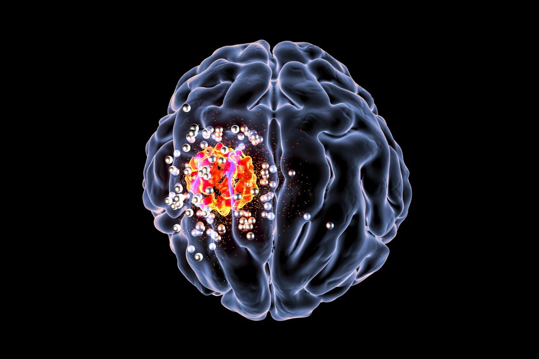
Glioblastoma Neuroimaging
Glioblastoma Neuroimaging Neuroimaging plays a central role in the initial diagnosis and subsequent monitoring of multimodal therapies offered to patients with glioblastoma – the most malignant type of brain cancer. The authors present a comprehensive description of current state-of-the-art clinical neuroimaging for glioblastoma. They cover the basic concepts and most recent applications of clinical structural neuroimaging, as well as the latest innovations and up-and-coming techniques for functional imaging of glioblastoma patients. Glioblastoma (GBM) is the most common adult primary intracranial
MRS imaging
Magnetic Resonance (MR) spectroscopy is a noninvasive diagnostic test for measuring biochemical changes in the brain, especially the presence of tumors. While magnetic resonance imaging (MRI) identifies the anatomical location of a tumor, MR spectroscopy compares the chemical composition of normal brain tissue with abnormal tumor tissue. This test can also be used to detect tissue changes in stroke and epilepsy. MR spectroscopy is conducted on the same machine as conventional MRI. The MRI scan uses a powerful magnet, radio
MRS Analysis
Equally as important as the pulse sequence used to record the data is the software used for data analysis. Ideally, fully automated spectral curve-fitting algorithms should be used to provide a quantitative estimate of each metabolite concentration. However, although such software exists and is often used in clinical research studies, it is little utilized in routine clinical practice, where visual interpretation (as in most radiological readings) is most common. Other commonly used metrics include ratios of metabolites such as NAA/Cho,

AI in Medical Imaging
One of the most promising areas of health innovation is the application of artificial intelligence AI in medical imaging, including, but not limited to, image processing and interpretation. Indeed, AI may find multiple applications, from image acquisition and processing to aided reporting, follow-up planning, data storage, data mining, and many others. Due to this wide range of applications, AI is expected to massively impact the radiologist’s daily life. Artificial intelligence (AI) can improve traditional medical imaging methods like Computed Tomography
تصویربرداری تومور مغزی
تومورهای مغزی یک مشکل مهم برای سلامتی در سراسر جهان هستند. به طور کلی ، آنها تومورهای بدخیمی هستند که بر روی انسان تأثیر می گذارند و در برابر تمام روشهای درمانی مقاوم میباشند. رگ زایی با تأمین اکسیژن و مواد مغذی برای حمایت از افزایش تکثیر سلولی و متابولیسم و حذف مواد زائد ، زمینه رشد کل تومور را فراهم می کند. با این حال هنگامی که تومورهای بدخیم بسیار کوچک هستند ، برای زنده ماندن اساساً به انتشار
تصویربرداری FMRI
تصویربرداری تشدید مغناطیسی عملکردی (تصویربرداری fMRI) تغییرات جزئی در جریان خون را که با فعالیت مغز اتفاق می افتد اندازه گیری می کند. ممکن است برای بررسی آناتومی عملکردی مغز (تعیین اینکه کدام قسمت از مغز عملکردهای مهم را انجام می دهند) ، ارزیابی اثرات سکته مغزی یا راهنمایی درمان مغز استفاده شود. تصویربرداری fMRI ممکن است ناهنجاری هایی را در مغز تشخیص دهد که با سایر روش های تصویربرداری یافت نمی شود. تصویربرداری fMRI در حال تبدیل شدن به