
تصویربرداری شناختی
تصویربرداری شناختی تصویربرداری شناختی از بسیاری جهات انقلابی در مطالعه علوم اعصاب شناختی ایجاد کرد ، رشته ای که سعی در تعیین مکانیسم های عصبی فرآیندهای شناختی دارد. مطالعات اولیه در مورد روابط مغز و رفتار ، به یک آزمایش دقیق عصبی متکی بود. ظهور تصویربرداری شناختی و ساختاری مغز ، ابتدا با توموگرافی رایانه ای و بعداً با تصویربرداری رزونانس مغناطیسی(ام ار ای) ، زمینه را برای محلی سازی دقیق آناتومیکی نقایص شناختی که پس از آسیب مغزی آشکار
تصویر برداری تومور مغزی
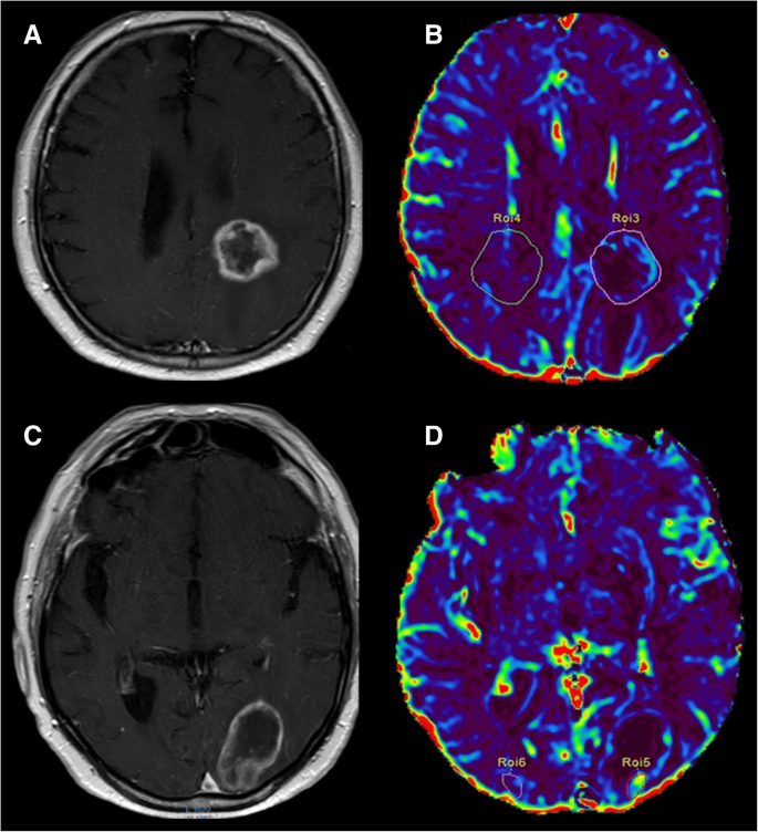
تومورهای مغزی یک مشکل مهم برای سلامتی در سراسر جهان هستند. به طور کلی ، آنها تومورهای بدخیمی هستند که بر روی انسان تأثیر می گذارند و در برابر تمام روشهای درمانی مقاوم میباشند. رگ زایی با تأمین اکسیژن و مواد مغذی برای حمایت از افزایش تکثیر سلولی و متابولیسم و حذف مواد زائد ، زمینه رشد کل تومور را فراهم می کند. با این حال هنگامی که تومورهای بدخیم بسیار کوچک هستند ، برای زنده ماندن اساساً به انتشار
نقشه برداری مغز
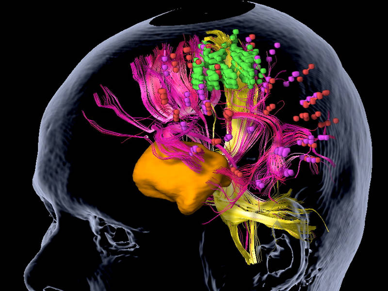
نقشه برداری مغز هنگام کار بر روی تومورهای مغزی ، جلوگیری از آسیب رساندن به نواحی مغزی که مسئول زبان ، حرکت و عملکرد حسی هستند بسیار مهم است. در حالی که می دانیم کدام قسمت های مغز مسئول این عملکردها هستند (و در کجاها واقعاً واقع شده اند) ، مغز هر فرد به اندازه کافی منحصر به فرد است که تغییرات جزئی وجود داشته باشد. بسته به نزدیک بودن تومور به هر یک از این نواحی ، ممکن است
تصویربرداری آلزایمر
تصویربرداری آلزایمر آلزایمر (زوال عقل) سندرم اکتسابی است که در آن فرد در یکی از شش حوزه شناختی زیر دچار اختلال پیشرونده می شود: توجه پیچیده ، عملکرد اجرایی ، یادگیری و حافظه ، زبان ، عملکرد ادراکی- حرکتی و / یا شناخت اجتماعی. این نقایص شناختی را نمی توان به اختلال روانی دیگری نسبت داد (به عنوان مثال ، افسردگی یا اسکیزوفرنی) و باید آنقدر شدید باشد که بتواند با عملکرد مستقل روزانه تداخل داشته باشد تا طبق نسخه

هوش مصنوعی در پزشکی
یکی از نوید بخش ترین زمینه های نوآوری در سلامتی ، تکامل هوش مصنوعی در پزشکی در زمینه تصویربرداری است که شامل پردازش و تفسیر تصویر است ، اما محدود به آن نیست. در واقع ، هوش مصنوعی در پزشکی ممکن است چندین برنامه را پیدا کند ، از دستیابی و پردازش تصویر گرفته تا گزارش دهی جهت کمک ، برنامه ریزی و پیگیری ، ذخیره داده و بسیاری برنامه های دیگر. با توجه به این طیف گسترده ای از
Texture analysis
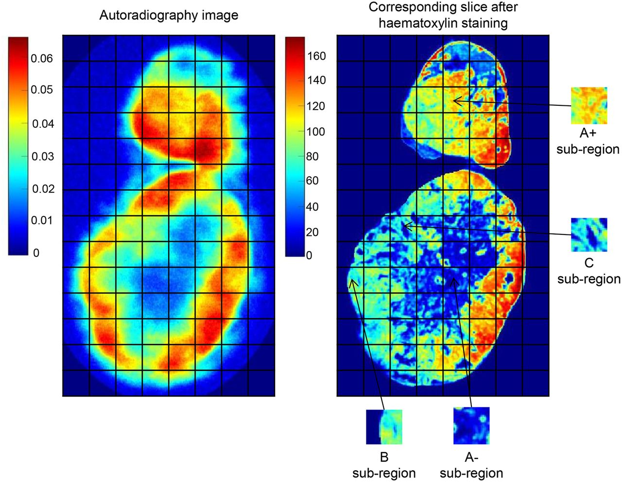
Description: Kio Medical has developed this app to quantify features related to the heterogeneity of the brain tissue and obtain texture parameters. Those texture parameters can be considered as indicators of tumor aggressiveness and help in brain tumor staging and phenotyping of pathological tissues. Disease: Solid tumors Glioblastoma Related Contents:
T2 Mapping
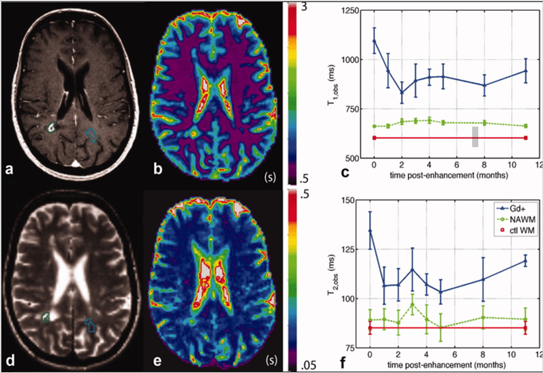
Description: Kio Medical has developed this app to automatically obtain the T2 mapping of tumors and depict changes and abnormalities with AI segmentation. The analysis of T2 mapping provides imaging biomarkers for the assessment and monitoring of solid tumors, such as prostate cancer, rectal cancer and liver cancer, among others. Disease: Solid tumors Prostate cancer Rectal cancer Endometrial cancer Liver cancer Related Contents:
rest state fMRI
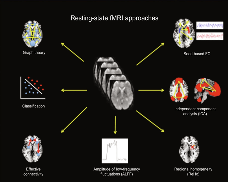
Description: Kio Medical has developed a software aimed at examining intrinsic networks in the brain while no task is performed to estimate correlations between brain regions. It measures spontaneous low-frequency fluctuations in the BOLD signal to investigate the functional architecture of the brain. One can either identify regions that have a close functional relationship with a given (seed) region, identify strongly connected regions, or perform a hypothesis-free independent component analysis across the whole brain. Its use in presurgical planning for

Diffusion Weighted Imaging (DWI) – IVIM
Description: Kio Medical’s software automatically performs intra-voxel incoherent motion (IVIM) analysis to detect changes in the microstructure of tissues to assess cancer processes in clinical practice and research. The tool allows the differentiation of pure diffusion changes due to variations in the vascular component and offers additional information complementary to conventional ADC results. This is useful for the characterization of tumoral processes to improve early detection, diagnosis, treatment response evaluation and follow-up of patients with gliomas, meningiomas and lymphomas. Disease:
Frontotemporal atrophy
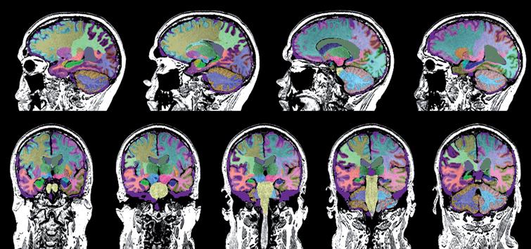
Description: Kio Medical has developed an app to quantify the volumes of the right and left hippocampus, frontal lobes and temporal lobes, to help assess disease diagnosis and progression. The obtained values are framed in a normative database to be compared with healthy subjects of the same age and gender. This software can be used to diagnose and monitor patients with Alzheimer’s disease, mild cognitive impairment and frontotemporal dementia. Disease: Alzheimer’s disease Mild cognitive impairment (MCI) Frontotemporal dementia Related